
SPIFFI microscopy breakthrough allows for real-time super-resolution imaging of living cells

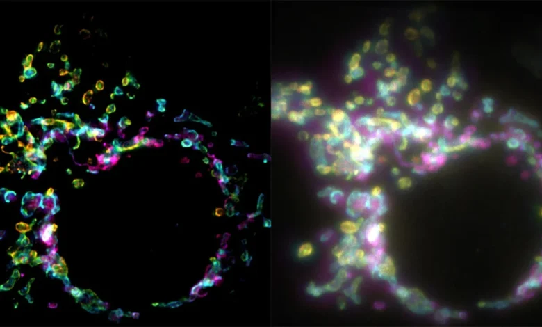
A pioneering leap in optical physics has emerged from the Laboratory of Nanoscale Biology (LBEN) at EPFL, where researchers have unveiled a novel fluorescence microscopy technique that overcomes one of the most persistent hurdles in modern cell biology: the trade-off between temporal resolution and spatial clarity. The new method, known as Spatial Polarization-induced Fluorescence Fluctuation Imaging (SPIFFI), enables the capture of super-resolution images from a single exposure. This development marks a fundamental shift from traditional techniques that require thousands of frames to reconstruct a single high-resolution image, a process that has historically rendered the study of rapid, live-cell dynamics nearly impossible.
The findings, published in the journal Nature, represent the culmination of years of inquiry into how the intrinsic properties of light—specifically polarization—can be harnessed to bypass the diffraction limit of conventional light microscopy. By shifting the paradigm from temporal data accumulation to spatial polarization analysis, the research team, led by Wei Guo, Lely Feletti, and Aleksandra Radenovic, has provided the scientific community with a tool that promises to turn the static, blurry images of the past into high-definition, real-time biological cinema.
The Diffraction Limit and the Evolution of Super-Resolution
To understand the magnitude of the SPIFFI development, one must first consider the "diffraction limit" established by Ernst Abbe in 1873. For over a century, scientists were restricted by the physical laws of light, which prevented conventional microscopes from resolving structures smaller than approximately half the wavelength of visible light—roughly 200 to 300 nanometers. In the cellular world, where essential structures like mitochondrial membranes, synaptic vesicles, and viral particles exist on a scale of tens of nanometers, this limit created a "blind spot" in our understanding of life at the molecular level.
The 21st century brought the Nobel-winning advent of super-resolution microscopy techniques such as STED (Stimulated Emission Depletion) and PALM/STORM (Photoactivated Localization Microscopy/Stochastic Optical Reconstruction Microscopy). These techniques successfully broke the diffraction barrier. However, they carried a significant caveat: they were inherently slow. Because they relied on the stochastic switching of individual fluorophores or the scanning of light beams across a sample, they required the collection of hundreds, or even thousands, of frames to produce one clear image.
In the time it took to capture these frames, a living cell—a dynamic, bustling environment—would have already changed shape, migrated, or altered its internal architecture. Consequently, these earlier methods were often relegated to fixed, dead samples, effectively turning "live" cell biology into a study of "frozen" snapshots.
The SPIFFI Mechanism: Leveraging Polarization
The SPIFFI technique, as detailed by Guo and his colleagues, pivots away from the temporal requirement. Instead of waiting for molecules to blink on and off over time, SPIFFI exploits the inherent polarization of fluorescent light. Fluorescent molecules, when excited, emit light that oscillates in specific directions based on their orientation within the cellular structure.
In a conventional setup, this information is often discarded or averaged out, leading to the "blurred" appearance seen in standard fluorescence imaging. SPIFFI, however, utilizes a sophisticated optical hardware configuration that splits this fluorescent light into four distinct, polarization-sensitive channels. By comparing these four images simultaneously, the system can mathematically resolve the orientation and position of molecules with high precision.
"Essentially, previous approaches used temporal information to resolve spatial resolution, but this doesn’t work very well on living cells," explains Wei Guo, the lead author of the study. Because SPIFFI captures all the necessary data in a single exposure, the temporal bottleneck is eliminated. The result is a system capable of producing high-fidelity, super-resolved frames at a speed limited only by the camera’s frame rate.
Experimental Validation and Technical Milestones
The research team conducted a series of rigorous experiments to validate the efficacy of the SPIFFI system. By targeting the mitochondrial outer membrane—a structure notoriously difficult to image due to its complex, constantly shifting morphology—the researchers demonstrated a two-fold improvement in resolution over standard widefield microscopy.
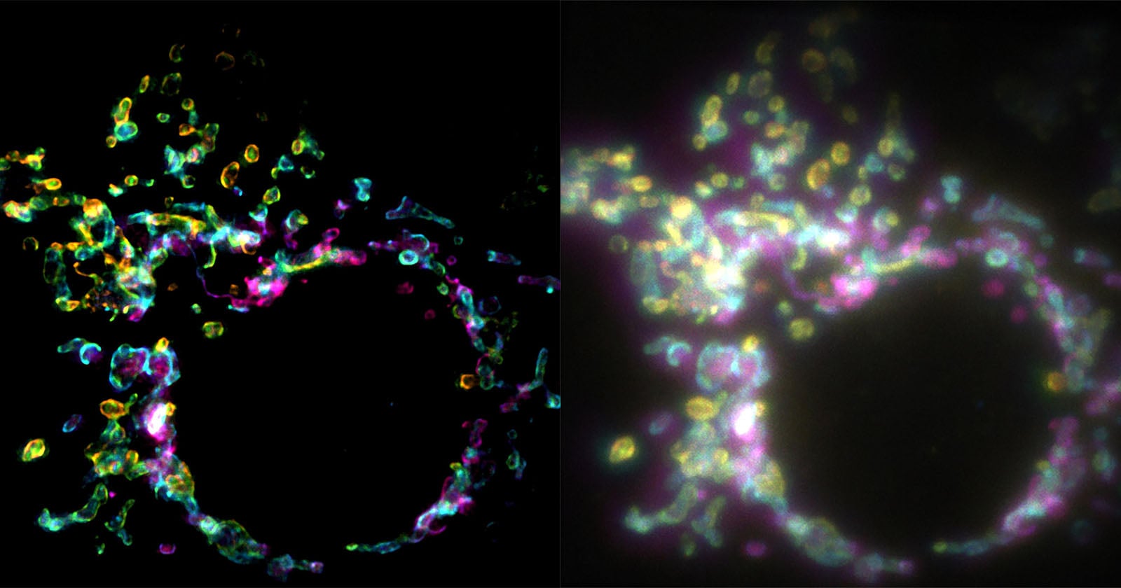
The experimental data indicates that SPIFFI can reliably resolve structures as small as 160 to 170 nanometers in a single frame. When the team integrated SPIFFI with existing fluctuation-based post-processing methods, they achieved resolutions of approximately 80 nanometers. This level of detail was previously reserved for imaging techniques that would have taken minutes to capture, whereas SPIFFI can generate these images in a fraction of a second.
Furthermore, the team successfully captured biological events that are notoriously difficult to track, such as cellular splitting (mitosis) and mitochondrial fusion. In these videos, the membrane structures remain crisp and defined, demonstrating that the technique can track the mechanical movements of organelles in real time without the "motion blur" that plagues slower, traditional super-resolution methods.
Implications for Life Sciences and Medical Research
The implications of this technology are vast. In neurology, for instance, understanding the rapid movement of vesicles within a synapse is critical to understanding how brain cells communicate. Traditional microscopy has provided only a rough estimation of these processes; SPIFFI offers the potential to visualize the actual docking and fusion of these vesicles as they occur.
In oncology, the technique could be used to monitor how cancer cells reorganize their internal infrastructure to migrate through tissues, potentially revealing new targets for therapeutic intervention. Because SPIFFI can be integrated into existing fluorescence microscopes—a significant upgrade in practicality compared to custom-built, multi-million dollar imaging suites—the barrier to entry for many research laboratories is significantly lower.
"With SPIFFI, every frame is super-resolved, meaning we can now produce super-resolution videos of live cells," Guo noted. This shift from "still photography" to "high-speed cinematography" at the nanoscale is expected to accelerate discoveries in developmental biology, virology, and pharmacology.
Future Outlook and Hardware Integration
As the researchers continue to refine the hardware, the primary goal is to increase the compactness and ease of use for the scientific community. The current SPIFFI hardware is designed to be modular, meaning it can be adapted to many existing commercial fluorescence platforms. This "add-on" philosophy is a departure from the industry trend of creating increasingly complex and proprietary imaging systems that require entire rooms of specialized equipment.
The EPFL team is now focused on optimizing the polarization-splitting optics to minimize light loss and improve signal-to-noise ratios in samples that are naturally dim. As the technology matures, it is likely that the principles of SPIFFI will be incorporated into the next generation of commercial biological microscopes.
While SPIFFI is not a replacement for every type of microscopy—it still faces the physical challenges of deep-tissue imaging and the inherent limitations of fluorescent labeling—it represents a vital missing link in the toolkit of the modern cell biologist. By decoupling spatial resolution from time, the researchers have effectively "freed" the cell from the constraints of the stopwatch.
As the scientific community begins to adopt these polarization-sensitive methodologies, the next few years are likely to see a surge in high-resolution, time-resolved studies that were previously thought impossible. From the movement of proteins within the crowded cytosol to the intricate, nanoscopic dances of organelles during cell division, the view inside the living cell has never been clearer, nor has it ever been captured with such remarkable temporal fidelity. The work of the LBEN team serves as a reminder that even in a field as mature as optics, there remain fundamental physical properties—like the polarization of light—that, when properly harnessed, can fundamentally change our perspective on the mechanics of life.
Related posts: