
Nikon Small World 2026 Photomicrography Competition Winners Reveal Microscopic Alien Worlds

Since its inception in 1975, the Nikon Small World Photomicrography Competition has served as a bridge between the clinical rigor of laboratory research and the evocative realm of visual art. Originally established to recognize excellence in light microscopy, the competition has evolved into a global benchmark for scientific imaging, showcasing the intersection of technical precision and aesthetic discovery. The 2026 results continue this tradition, highlighting a collection of images that transform mundane biological structures and mechanical processes into landscapes that appear almost extraterrestrial.
The Evolution of a Scientific Showcase
When Nikon launched this initiative over five decades ago, the primary objective was to celebrate the work of researchers and enthusiasts who utilized optical microscopes to document the invisible. At the time, such imagery was largely confined to peer-reviewed journals and academic textbooks, often lacking the public visibility afforded to other forms of nature photography. Over the years, the competition has expanded its scope, inviting participants from diverse backgrounds—including biologists, engineers, and hobbyist photographers—to submit entries that demonstrate not only technical mastery but also a creative perspective on the microscopic scale.
The 2026 iteration attracted thousands of entries from across the globe, each scrutinized by a panel of judges comprising experts in both microscopy and professional photography. The criteria for success are stringent: entrants must balance scientific accuracy with composition, lighting, and clarity, often requiring hours of preparation for a single frame.
A Technical Triumph: The 2026 Winning Entry
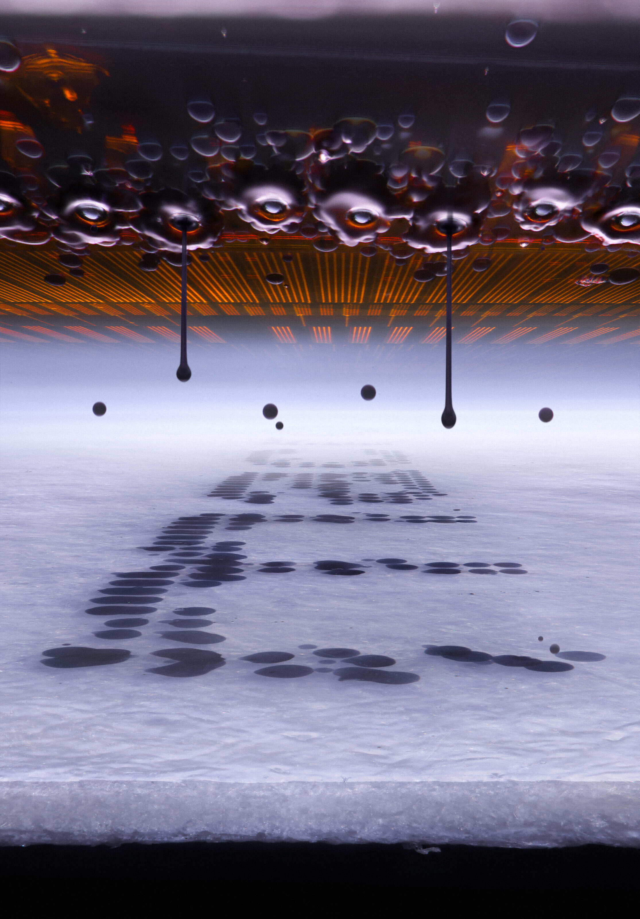
The top honor for 2026 was awarded to Ole Bielfeldt, a 25-year-old design student from Germany. His winning submission captures a moment of mechanical precision: the instant black ink is expelled from an inkjet printer nozzle onto paper. The image, characterized by its sharp focus on the fluid dynamics of the ink, represents the culmination of a rigorous, DIY-oriented process.
Bielfeldt’s approach deviates from traditional laboratory methodology. Rather than utilizing pre-manufactured, high-end equipment exclusively, he focuses on bespoke, modified setups. By experimenting with lighting and timing, Bielfeldt managed to visualize the chaotic yet structured nature of inkjet deposition. "The process of iteration is where the real work happens," Bielfeldt noted in a statement regarding his success. "Failures are an integral part of the discovery phase. When you are looking at the world through a lens at this magnification, you have to embrace the unpredictability of the subject matter."
The Intersection of Biology and Aesthetics
While the top prize went to a mechanical subject, the remainder of the top-ranked entries underscored the complexity of biological life. The competition frequently highlights structures that are essential to human and animal life but are rarely seen by the naked eye.
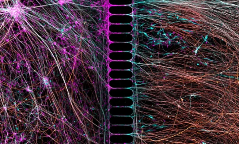
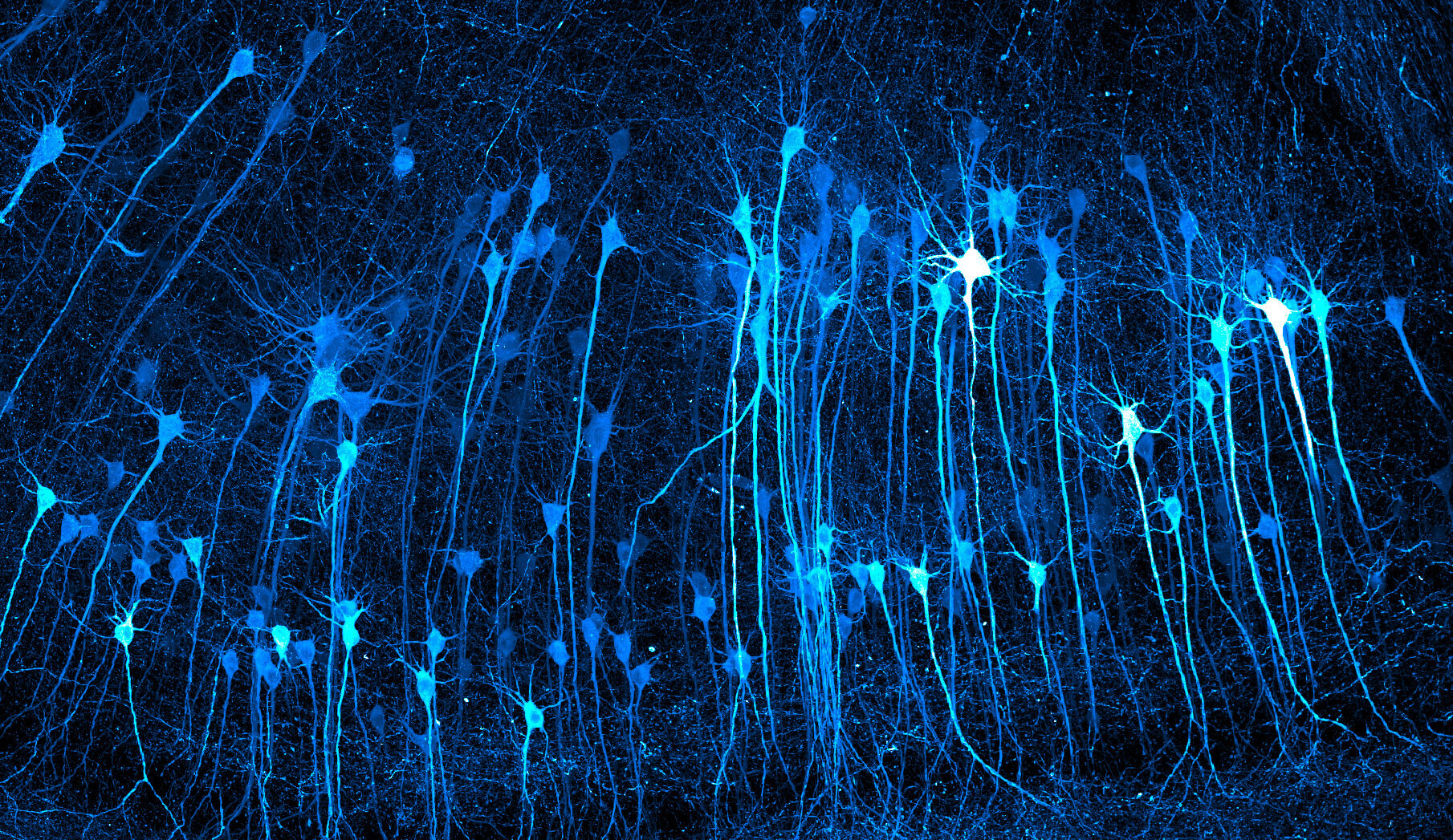
Third place was secured by Kaelin Wulf and Rachel Flannery for their 5X-magnified image of neurons within a mouse brain. The image provides a high-contrast visualization of neural pathways, emphasizing the density and interconnectivity of the central nervous system. Such images are not merely artistic; they are vital tools for researchers studying neurodegenerative diseases. By mapping these pathways in detail, scientists can better understand the physical manifestations of complex cognitive disorders.
Similarly, Dr. Bruno Cisterna and Dr. Eric Vitriol from the Medical College of Georgia at Augusta University earned the 7th-place position for their work on human neurons. Utilizing confocal imaging—a technique that allows for the creation of high-resolution, three-dimensional optical sections—they photographed neurons grown from reprogrammed skin cells. This methodology, known as induced pluripotent stem cell (iPSC) technology, is a cornerstone of modern regenerative medicine. The ability to visualize these cells in such vivid detail allows researchers to monitor the efficacy of new drug therapies and cellular interventions before they reach clinical trials.
The "Martyrs" of Science: Mouse Anatomy
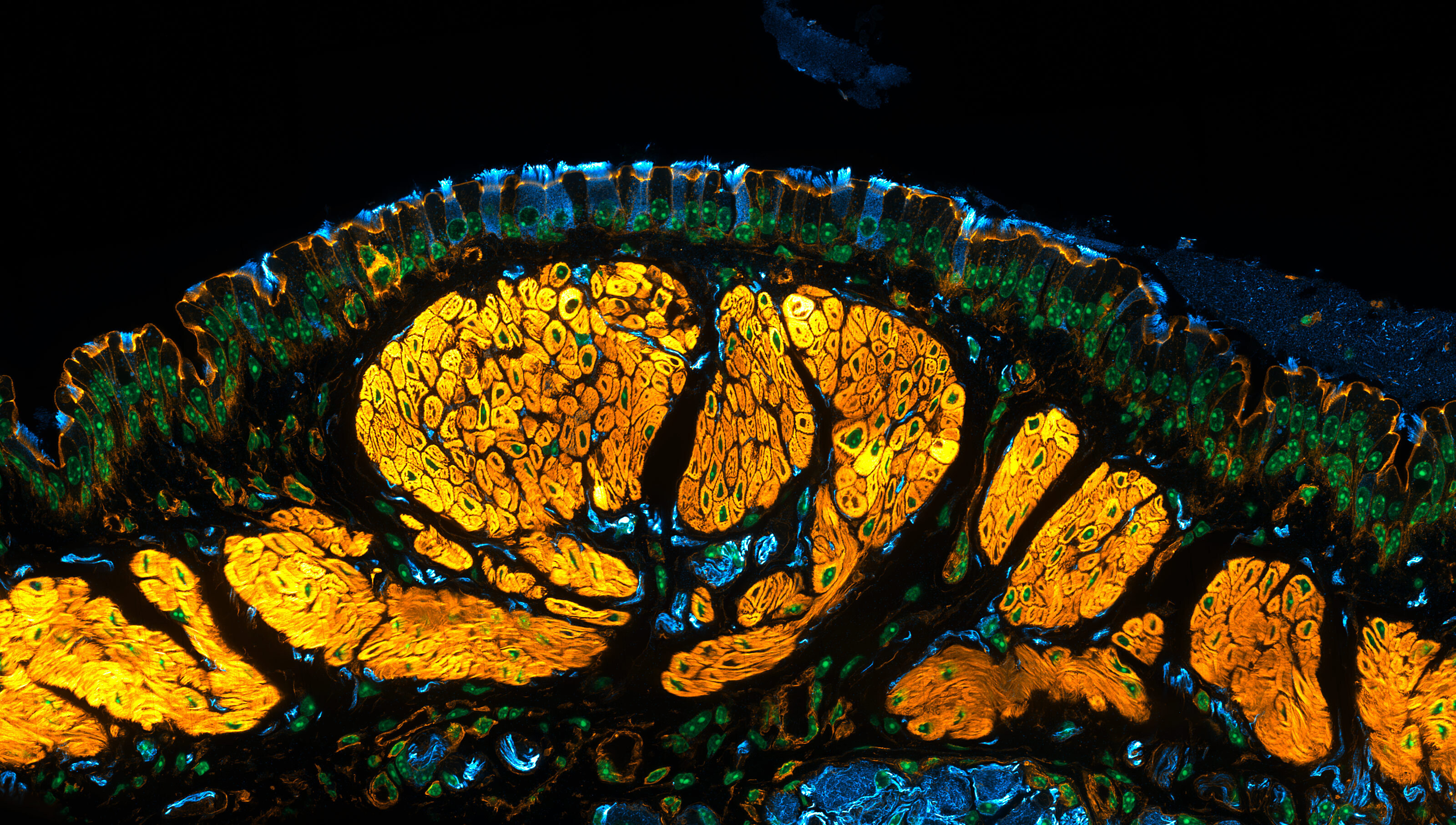
Mice occupy a paradoxical position in the history of science. They are the most frequently used model organisms in biomedical research, yet they are rarely viewed through an artistic lens. The 2026 competition featured several entries that elevated mouse anatomy to high art, including a 15th-place entry by Dr. Julien Resseguier, which depicted a mouse trachea at 60X magnification. The image, which features intricate, stained-glass-like structures, demonstrates how histological preparations—often treated with dyes to highlight specific proteins or tissues—can yield results that are visually arresting.
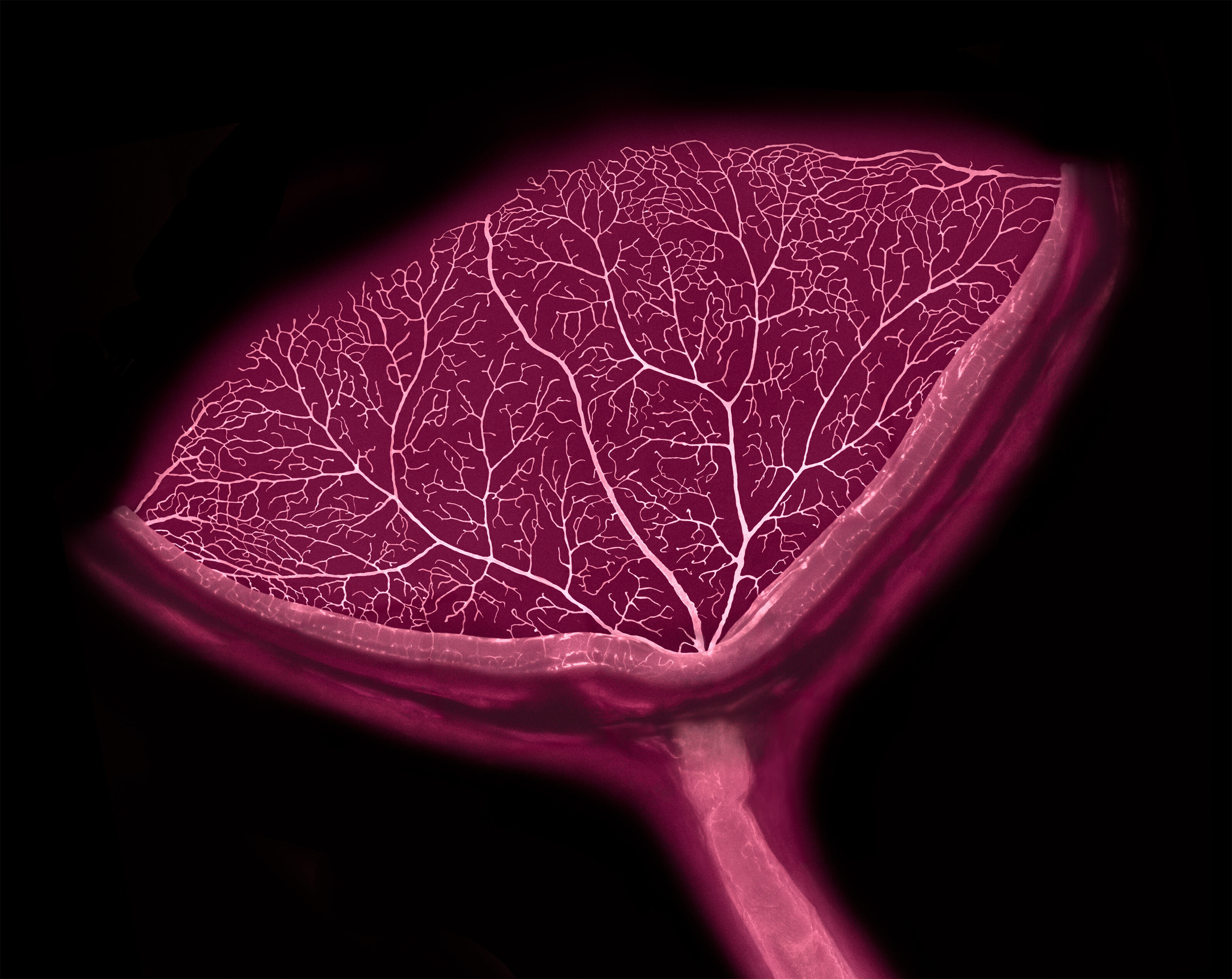
Additionally, Satu Paavonsalo and Dr. Sinem Karaman from the University of Helsinki provided a detailed look at a mouse’s eye, capturing the optic nerve and surrounding blood vessels. This image, which secured 19th place, was originally produced during the course of a drug therapy study. By highlighting the vascular network of the eye, the researchers were able to assess the impact of their therapeutic agents on ocular health.
The Artistic Edge: Nature’s Surrealism
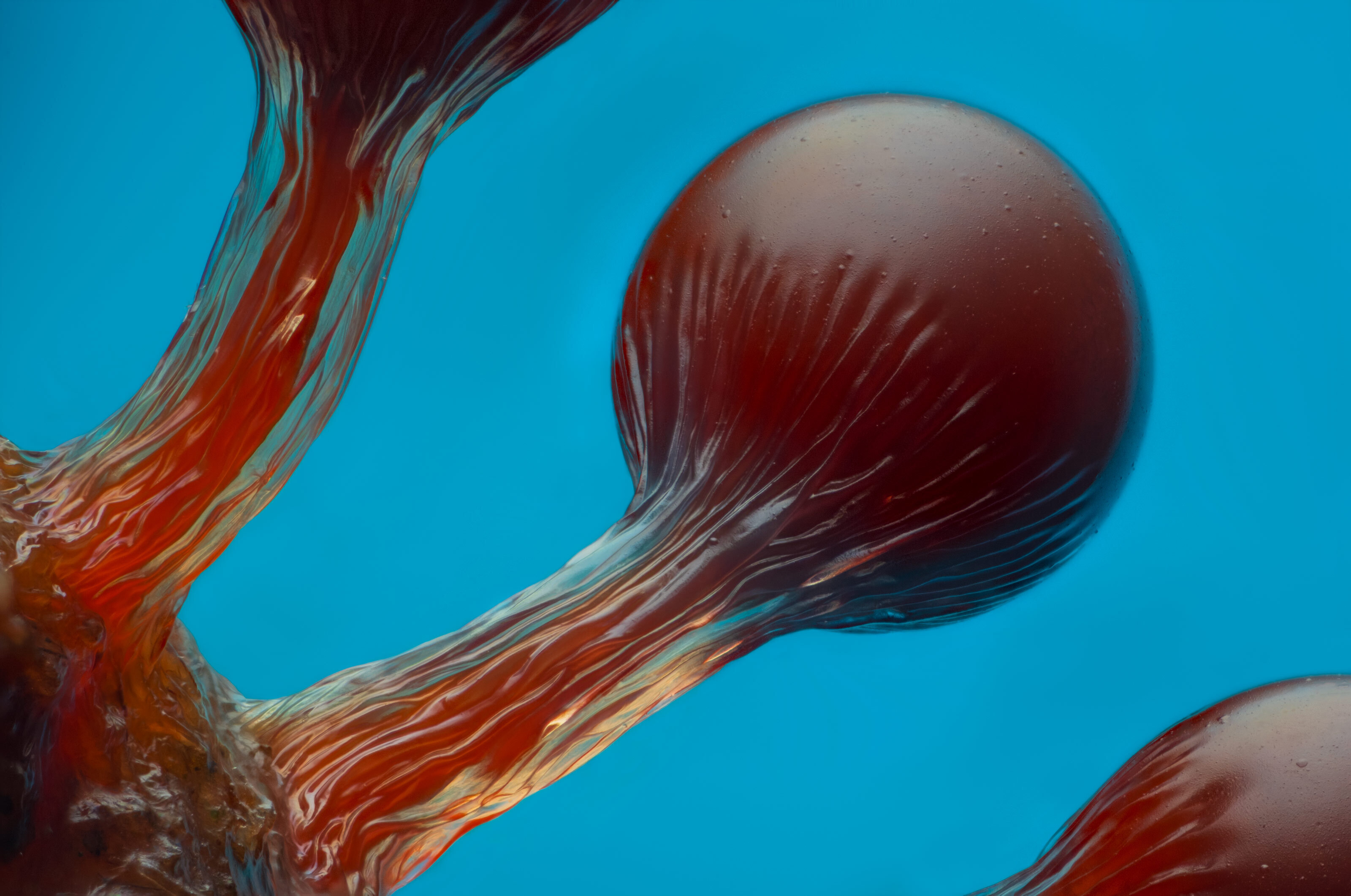
The competition also celebrates the diversity of the natural world, as seen in the 6th-place entry by Eric Epstein, which featured the fruiting bodies of Hemitrichia slime mold. The image’s vibrant colors and whimsical, bulbous forms drew comparisons to the surrealist landscapes found in literature, illustrating that even organisms traditionally dismissed as simple biological growth possess a distinct, alien architecture.
Second-place winner John-Oliver Dum offered a different perspective with his close-up of a jumping spider. The subject, covered in grains of pollen cultivated by his wife, serves as a reminder of the symbiotic relationships between insects and flora. Dum’s fascination with arachnids, rooted in childhood experiences, translates into a portrait that challenges the viewer to reconsider their perceptions of these common, yet often feared, creatures.
Broader Implications and Scientific Communication
The impact of the Nikon Small World competition extends beyond the photography community. In an era where scientific literacy is increasingly critical, these images serve as a vital medium for public engagement. By translating complex biological functions into accessible, visually stunning images, the competition helps demystify scientific research.
Furthermore, the data produced by these photographers often informs subsequent academic inquiry. The "Small World" archive now contains decades of visual data that reflect the evolution of microscopy technology—from early light-based optics to current confocal and fluorescence scanning techniques. As imaging technology becomes more accessible, the barriers to entry for such high-quality documentation are lowering, allowing for a broader cross-section of global talent to contribute to the scientific record.
Future Outlook
As the competition looks toward 2027 and beyond, the trend of integrating DIY engineering with professional scientific research is expected to accelerate. The success of winners like Bielfeldt suggests that the future of photomicrography lies in the hands of those who are willing to bridge the gap between traditional laboratory equipment and creative, improvised technical approaches.
For the scientific community, these images are a testament to the fact that the most profound discoveries often occur at the edges of our perception. Whether it is the study of human neurons or the fluid dynamics of a common office printer, the microscopic world remains a frontier of both technical challenge and immense beauty. As these winners continue to showcase, the ability to see the world at a scale of micrometers is not just a requirement for discovery—it is an opportunity to view our reality with a renewed sense of wonder.
The full gallery of the 2026 Nikon Small World competition, including the honorable mentions and technical specifications for each image, remains accessible to the public, providing a comprehensive resource for educators, students, and researchers alike.